

Diagnostic radiology uses medical imaging to detect and evaluate conditions throughout the body. Neuroradiology is a subspecialty focused on the brain, spine, and nervous system; providers use techniques like MRI and CT scans to identify structural abnormalities. Epilepsy is a neurological disorder marked by recurrent, unprovoked seizures caused by abnormal electrical activity in the brain. When evaluating epilepsy, physicians turn to medical imaging to locate structural changes that may be triggering seizures, and neuroradiology provides the tools to do exactly that.
Causes of Epilepsy
Epilepsy can stem from a wide range of structural, genetic, and acquired factors. Brain conditions such as tumors, strokes, and cortical dysplasia are among the recognized structural causes, and head injuries or infections like meningitis can also trigger the disorder. When no identifiable cause is found, the condition is classified as idiopathic.
Genetic factors play a role in several epilepsy syndromes, and researchers have identified specific gene mutations that affect how neurons transmit signals. The cause often determines the type of seizure a person experiences, which shapes how physicians approach evaluation and management. Neuroradiology helps diagnose epilepsy and conduct imaging studies of the nervous system.
Symptoms of Epilepsy
Epilepsy symptoms vary widely, since the type of seizure depends on where in the brain abnormal activity begins. Generalized seizures affect both hemispheres simultaneously, but focal seizures originate in one specific brain region. Both types produce distinct presentations.
Common symptoms include:
- Sudden, uncontrolled convulsions or muscle stiffness
- Brief episodes of staring or loss of awareness
- Temporary confusion or memory lapses
- Sensory changes such as tingling, visual disturbances, or unusual smells
- Sudden falls without a clear cause
Diagnosis of Epilepsy
Diagnosis begins with a thorough clinical history and neurological examination. Because no single test confirms epilepsy, physicians combine several approaches to build an accurate picture. An electroencephalogram, or EEG, records the brain’s electrical activity, and abnormal patterns can support a diagnosis of epilepsy.
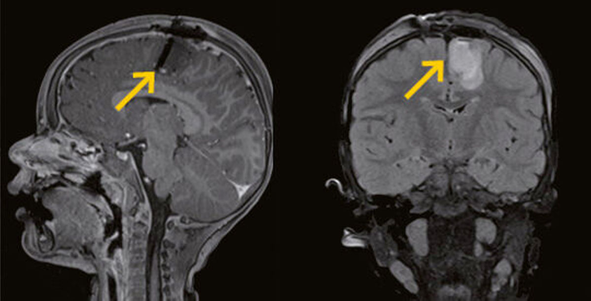
Neuroimaging plays a role in identifying structural causes. MRI is the preferred imaging modality; it offers high-resolution views of brain tissue and can detect abnormalities such as hippocampal sclerosis, malformations of cortical development, and low-grade tumors. Advanced MRI techniques, including functional MRI (fMRI) and diffusion tensor imaging, or DTI, allow clinicians to map brain activity and white matter pathways, which aids in pre-surgical planning for patients with drug-resistant epilepsy. PET scans and SPECT imaging may supplement MRI by revealing metabolic changes in brain regions associated with seizure onset.
Signs of Epilepsy
Seeking medical evaluation promptly can make a meaningful difference in outcomes for people with epilepsy. Some seizures can resemble simply “zoning out” for a period of time. Consult a doctor if you experience:
- A first-time seizure of any kind
- Seizures that last longer than five minutes
- Repeated seizures without regaining full consciousness in between
- Seizures accompanied by fever, head injury, or pregnancy
- Prolonged confusion following a seizure episode
Even a single unexplained seizure deserves evaluation. Some seizures can indicate an underlying structural problem that requires prompt imaging, and early detection of a cause can guide effective treatment.
Learn More About Neuroradiology
Neuroradiology continues to advance the way physicians evaluate epilepsy. Modern imaging identifies structural abnormalities in the brain and supports surgical planning; these technologies help distinguish epilepsy from other neurological conditions. To learn more about neuroradiology, consult a radiology imaging clinic near you.