

An echocardiogram is a non-invasive imaging test that uses sound waves to create detailed pictures of the heart. It is a widely used diagnostic tool in cardiology; healthcare providers rely on it to study the heart’s structure and function. Knowing what the test involves and what it may reveal enables you to have more productive conversations with your care team. Here’s information about this procedure and how it helps diagnose heart conditions:
Understanding Echocardiogram Basics
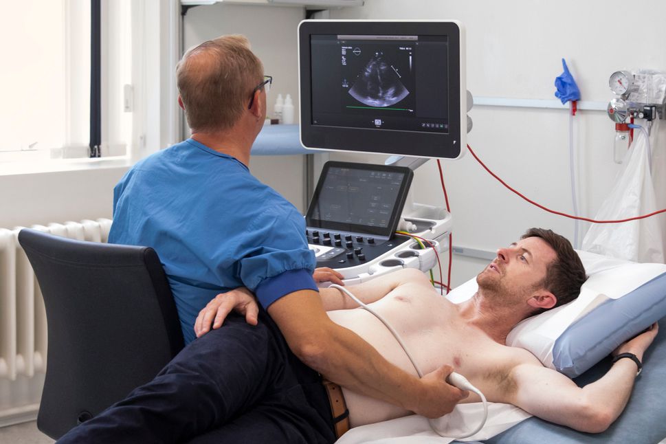
An echocardiogram is aspecialized type of ultrasound, often used for diagnosing heart problems. A device called a transducer sends high-frequency sound waves toward the heart, and those waves bounce back to form moving images on a monitor. The test is painless and does not involve radiation.
Providers may use several types of echocardiography. A standard transthoracic echocardiogram places the transducer on the chest, while a transesophageal echocardiogram passes the transducer down the throat for closer images. Your provider selects the most appropriate option for your situation; this decision depends on the detail required, your symptoms, and the heart area being examined.
Evaluating Key Heart Functions
Echocardiograms give providers a real-time look at how the heart performs, allowing them to assess several key areas during the test. Providers can examine heart structure and chamber size. They study the shape of the heart’s four chambers; enlarged chambers or thickened walls sometimes indicate conditions worth investigating further.
Providers assess valve movement. The heart has four valves that regulate blood flow, and an echo shows how well each one opens, closes, and directs blood in the correct direction. They can also evaluate blood flow patterns using Doppler imaging to identify any abnormal flow or leakage.
The pumping function is another fundamental focus. One key measurement, the ejection fraction, reflects how much blood the left ventricle pushes out with each heartbeat. This figure helps providers gauge overall pumping efficiency.
Supporting a Broader Assessment
An echocardiogram produces helpful data, yet it remains one piece of a larger diagnostic picture. Providers evaluate the results alongside your medical history, physical examination findings, and other tests such as ECGs or blood work. The results help providers identify potential structural or functional concerns. They assist with narrowing down possibilities and determining the next steps. Those steps may include additional imaging, specialist referrals, or adjustments to a monitoring plan.
Understanding Reasons for the Procedure
Providers recommend an echocardiogram for a range of reasons:
- Investigating unexplained symptoms such as shortness of breath or an irregular heartbeat
- Monitoring a previously identified heart condition over time
- Evaluating heart function before or after a procedure
- Assessing structural abnormalities detected during a physical exam
- Checking how well the heart responded to a treatment plan
Your provider will supply specific pre-test instructions based on the type of echocardiogram ordered. A standard transthoracic echocardiogram typically requires no special preparation; other types may involve dietary restrictions or medication adjustments prior to the procedure. Patients are advised to wear comfortable, loose-fitting clothing. Bring any relevant medical records along with an up-to-date list of current medications to the appointment. These preparatory measures help promote an efficient and well-coordinated clinical visit.
Learn More About Echocardiograms
An echocardiogram is a practical, well-established tool that shows providers a detailed view of the heart’s structure and function. It plays a key role across a wide range of cardiac assessments; this includes evaluating valve performance and assessing overall cardiac pumping efficiency. Consult your care team to learn more about this procedure.