
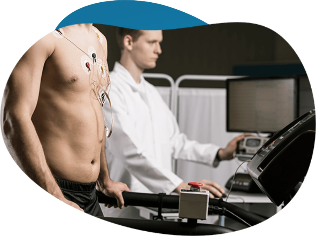

Nuclear stress testing gives doctors a detailed picture of how the heart performs under pressure, and it helps doctors guide treatment decisions. For patients scheduled to undergo this procedure, understanding each step can make preparation easier. Here is more information on what nuclear stress testing is:
What Is Nuclear Stress Testing?
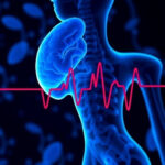
Nuclear stress testing is a diagnostic imaging procedure used to evaluate blood flow to the heart muscle. It combines a standard stress test with nuclear imaging, allowing cardiologists to detect areas of the heart that may not be receiving adequate blood supply. The test produces detailed images of the heart both at rest and under stress conditions, and consulting a healthcare professional helps determine when the test is needed.
How Does It Work?
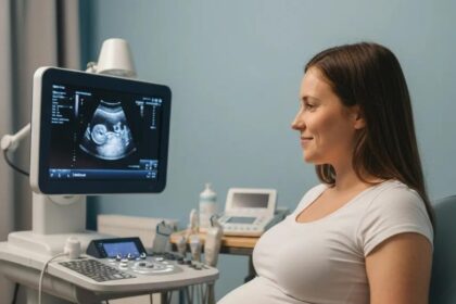
A small amount of radioactive tracer is injected into the bloodstream, and it then travels to the heart muscle. A specialized gamma camera captures images of the tracer’s distribution. It highlights areas with normal and reduced blood flow. Two sets of images are typically taken, including one while the heart is at rest and one after it has been stressed.
The stress phase can be induced through physical exercise on a treadmill or stationary bike, or through a pharmacological agent if the patient is unable to exercise. Pharmacological agents dilate the coronary arteries, and it simulates the effects of exercise. The imaging process may take several hours.
Why Is It Conducted?
Cardiologists order these tests to evaluate coronary artery disease, a condition where narrowed arteries reduce blood flow to the heart. The test helps assess the extent of damage after a heart attack, and it measures how well the heart pumps blood. It guides decisions about procedures such as angioplasty or bypass surgery. Patients experiencing chest pain, shortness of breath, or unexplained fatigue may be referred for this test.
What Happens Before and After?
Before the test, patients may be asked to:
- Avoid eating or drinking hours beforehand
- Refrain from consuming caffeine
- Discuss current medications with their physician
After the resting images are taken, the stress portion of the test begins. Following this, a second round of imaging captures changes in blood flow under stress. Patients may feel flushed, short of breath, or experience mild chest discomfort during the pharmacological phase; these sensations are temporary and expected.
When the test is concluded, patients can return to normal activities relatively quickly. The radioactive tracer clears from the body naturally, primarily through urine. A healthcare professional will analyze the images, and results may be shared with the referring physician within a few days.
What Are the Benefits?
This testing offers a non-invasive way to assess cardiac health with a high degree of accuracy. It can detect coronary artery disease at an early stage, before symptoms become severe, allowing for timely intervention. The detailed imaging it produces gives physicians more actionable information than a standard exercise stress test alone, and it guides treatment planning.
The radiation exposure from the tracer is minimal and safe for most patients. Since the procedure combines functional and imaging data, it can reduce the need for more invasive diagnostic procedures. Learning more about the test helps patients prepare, and speaking with a healthcare professional is valuable.
Know What To Expect
Nuclear stress testing is a procedure that provides doctors with detailed information about heart function. Knowing what each stage involves allows patients to prepare for the process. If your physician has recommended this test, follow their pre-procedure instructions carefully and ask any questions you have before the appointment.