

An echocardiogram, which uses high-frequency sound waves, is a non-invasive cardiac imaging test that provides detailed images of the heart’s structure and function. Doctors may recommend this procedure to evaluate symptoms such as chest pain, shortness of breath, or irregular heartbeat, and it is also used to monitor existing cardiac conditions. Understanding what the procedure involves helps patients make informed decisions.
What Is an Echocardiogram?
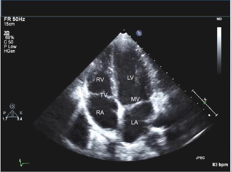
An echocardiogram is an ultrasound of the heart. It uses high-frequency sound waves, which produce real-time images of the heart’s chambers, valves, walls, and connected blood vessels. These images allow cardiologists to assess cardiac function, and they can also determine whether the heart’s structures appear normal.
Several types of echocardiograms are available, each of which serves a distinct clinical purpose depending on the information needed. The transthoracic echocardiogram (TTE) is the standard type; it is performed externally on the chest. A stress echocardiogram combines standard echocardiography with physical or pharmacological stress to evaluate how the heart functions under exertion. Doppler echocardiograms use Doppler ultrasound technology to measure blood flow speed and direction through the heart’s chambers and valves.
Why Is It Conducted?
A cardiologist may order an echocardiogram for many reasons, and the specific reason depends on the patient’s clinical presentation. Common reasons include evaluating symptoms such as chest pain, shortness of breath, or heart palpitations. Monitoring known conditions like heart failure, valve disease, or congenital heart defects is another indication. The test measures the heart’s ejection fraction, which reflects how effectively the heart pumps blood with each beat. A low ejection fraction can signal weakened heart muscle function, and this finding guides subsequent treatment decisions.
What Does the Process Involve?
Before a standard TTE, no special preparation is typically required. The patient may be asked to lie on an examination table, where a sonographer will apply a small amount of gel to the chest. A sonographer then moves a handheld transducer across the skin, and they capture detailed images of the heart’s structures.
The procedure is painless, and it generally takes about 60 minutes. Once the test is complete, a cardiologist reviews the images and analyzes the report. Some patients may be asked to undergo a stress echocardiogram, which incorporates a physical or pharmacological stress component. This variation lets doctors see how the heart performs under increased demand.
What Are the Benefits?
An echocardiogram offers a non-invasive, radiation-free way to evaluate cardiac health. The real-time imaging capability of an echocardiogram is particularly valuable:
- It allows cardiologists to observe the heart in motion, not just in a static state
- It can detect abnormalities in wall movement, valve function, and fluid accumulation around the heart
- It enables immediate assessment of cardiac response during stress testing or other diagnostic procedures
Early detection of structural or functional abnormalities can lead to more timely and targeted treatment decisions.
Know What To Expect
An echocardiogram is a low-risk procedure that gives cardiologists a detailed view of how your heart is functioning. From identifying valve abnormalities to measuring pumping efficiency, the information gathered through this test informs your care plan, and no lengthy recovery time is needed. If you have been referred for an echocardiogram, ask your cardiologist what to expect.