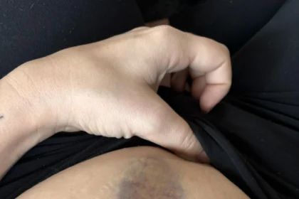


The skin functions as the body’s largest organ and acts as a primary defense against environmental elements. Throughout life, various spots, freckles, and marks naturally appear across the surface. While the vast majority of these growths remain entirely harmless, observing the skin helps detect structural variations early on. A skin doctor evaluates skin lesions to differentiate between normal benign growths and atypical moles that might require laboratory testing. Understanding what makes a mark irregular aids in recognizing when a professional examination is warranted.
What Is an Unusual Mole?
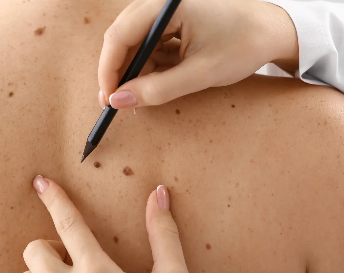
Recognizing atypical spots requires a systematic and structured approach. A skin doctor may recommend the ABCDE method to assess skin lesions accurately. ‘A’ represents asymmetry, meaning one half of the spot looks visibly different from the exact opposite half. ‘B’ stands for border irregularity and presents as blurred or jagged edges rather than a smooth circle. ‘C’ denotes color variation, where a single spot displays multiple mixed shades. ‘D’ refers to diameter, specifically noting spots larger than six millimeters across. ‘E’ represents evolution, marking any shifts in shape, size, color, or physical texture over time.
Other noticeable signs include a completely new spot appearing during adulthood or a dark streak developing underneath a fingernail or toenail bed. Sudden, unexpected bleeding from a mole is another symptom, or persistent itching from a mark can also occur. Any fast-growing pink or red bump also necessitates a prompt professional medical review, as some abnormal growths entirely lack dark pigmentation.
What Causes Them?
A primary factor driving abnormal skin cell growth is exposure to ultraviolet radiation. This includes recurrent direct sun exposure and specific instances of severe sunburns. Indoor tanning beds deliver highly concentrated ultraviolet rays and raise the likelihood of abnormal cellular changes. Specific physical traits frequently link to higher risk profiles, including fair skin, light-colored eyes, and blonde or red hair. A high volume of standard moles across the body, a distinct family history of skin irregularities, or a weakened immune system also elevate the likelihood of developing abnormal lesions. Because some irregularities emerge in areas completely shielded from sunlight, full-body observations remain a necessary preventive practice.
How Are They Treated?
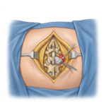
A clinical visit typically starts with a thorough full-body examination from the scalp down to the feet. The skin doctor uses a specialized magnification device, known as a dermatoscope, to view microscopic structures that remain invisible to the naked eye. If a specific lesion displays concerning characteristics, the physician may perform a minor in-office procedure called a biopsy. This process involves numbing the immediate surrounding area with a local anesthetic and removing a small tissue sample for laboratory analysis.
Laboratory results directly guide the subsequent clinical steps. If the tissue proves benign, no further intervention is typically needed. For severely malignant findings, the doctor proceeds with a local excision. This surgical technique removes the problematic tissue alongside a measured perimeter of healthy skin; this helps verify that no abnormal cells remain in the body.
Meet With a Skin Doctor
Scheduling a skin examination establishes a definitive reference point for your overall skin health. During this initial visit, a dermatologist can formulate a specific follow-up schedule for you and base it on your individual risk factors. Between clinical visits, conducting a thorough monthly self-examination using a full-length mirror and a handheld mirror helps track any visible physical shifts.