

Magnetic resonance imaging (MRI) uses magnetic fields and radio waves to produce detailed images, and doctors use these scans to view organs, tissues, and bones. While the technology is a standard tool, recent developments have refined how MRI machines work. Here are some advancements in MRI imaging technology:
High-resolution Imaging
Modern MRI systems capture images with higher resolution than older models. Engineers improved the magnets and software, and these upgrades produce sharper results. Since the detail is clearer, radiologists may read scans with greater accuracy. New scanners also work faster than past versions.
Faster scans typically mean shorter appointment times for patients, and while speed matters, image quality remains the priority for most clinical teams. Stronger magnets generate finer detail, and updated coils support that process. These tools help physicians spot small changes in the body.
Wide-bore MRI Technology
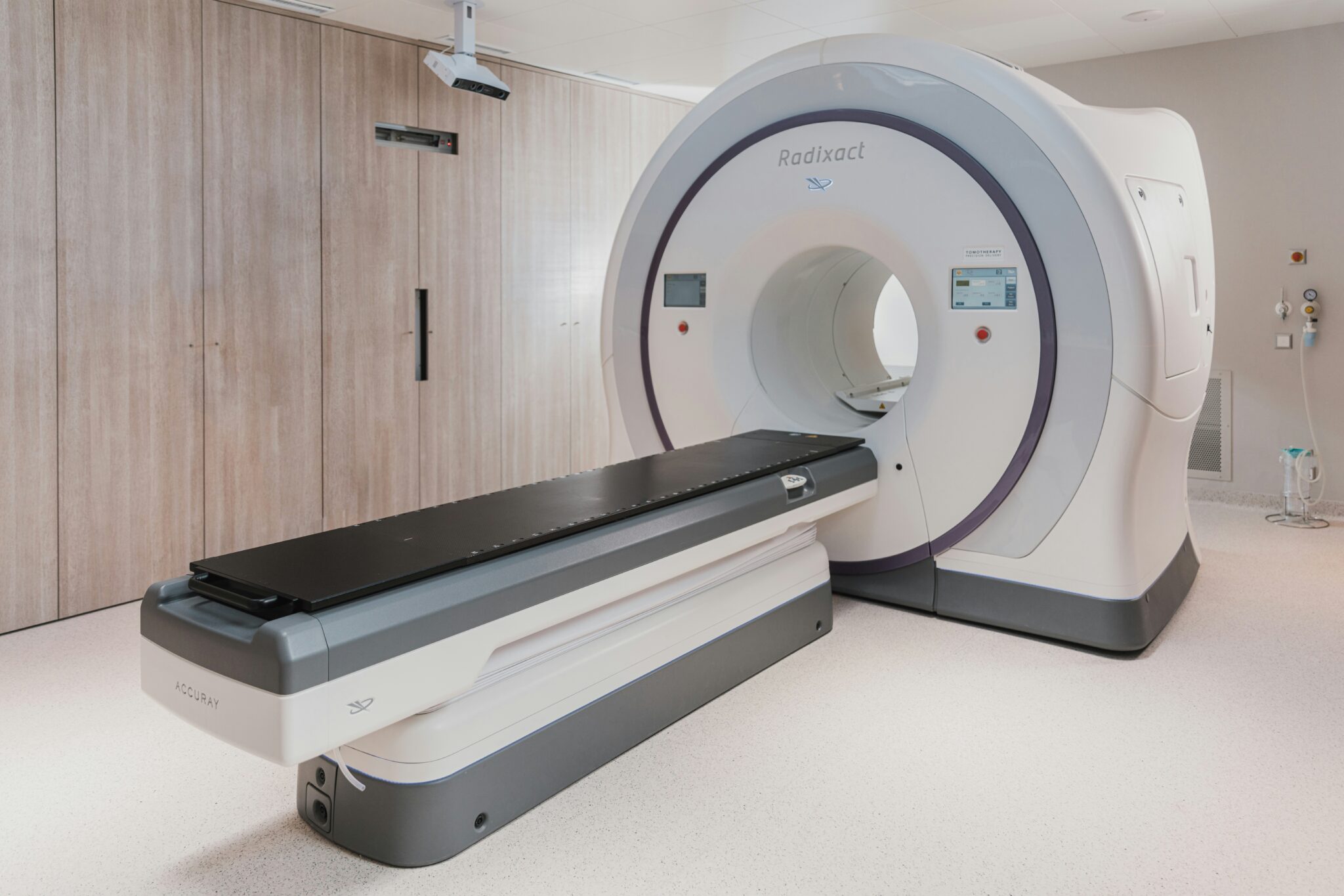
A wide-bore MRI features a larger opening than a standard machine. The diameter is typically measured wider. Since the tunnel is wider, the scans are less confined. Many facilities now install these units to serve more patients.
Wide-bore designs suit several specific groups, and clinics note their practical value:
- People with larger body types
- Patients who avoid tight spaces
- Individuals recovering from certain injuries
As the bore is wider, the imaging strength stays strong. Technicians position patients comfortably, and the scan proceeds as planned. The wider opening changes the experience without lowering image quality.
Increased Space
Extra room inside the machine addresses a typical concern. Some patients have trouble in narrow tunnels, so manufacturers expanded the interior. Since the space is larger, people typically keep still during the scan.
Stillness affects the final images directly. When a patient moves, the scan may blur and need to be repeated. The added space helps reduce that risk, and it supports a smoother session. Clear images come from steady positioning.
Some newer machines also shorten the length of the tunnel. A shorter tunnel lets parts of the body remain outside during certain scans. Since more of the body stays exposed to open air, the experience may improve. Facilities choose these models for varied patient needs.
Soft-tissue Imaging
MRI excels at showing soft tissues that other scans miss. The contrast between muscle, fat, and organs appears clearly. Since no radiation is involved, MRI differs from X-rays and CT scans, and this feature makes it useful for many diagnoses.
Doctors use soft-tissue imaging to examine specific structures, and the list below shows major targets:
- Brain and spinal cord
- Ligaments and tendons
- Organs
Detailed views help guide treatment decisions. When a physician sees tissue clearly, the diagnosis may follow more directly. Soft-tissue contrast remains one of the main reasons doctors order MRI scans. The technology continues to support that role.
Schedule an MRI Near You
If your doctor recommends a scan, ask about the equipment available nearby. Knowing your options helps you prepare for the appointment, and a quick phone call answers many of your questions. Contact a local imaging center today to schedule your MRI, speak with the staff about your needs, and confirm the type of machine they use. Reach out now and take the next step toward your scan.